Chest Muscles Anatomy Labeled - Muscle Diagram Most Important Muscles Of An Athletic Black Man Anterior And Posterior View Male Body Labeled Vector Illustration Chart On White Background Premium Vector In Adobe Illustrator Ai Ai / Male shoulder and chest muscles labeled chart on white labeled human anatomy diagram of male shoulder, biceps, arm, and chest muscles frontal anterior view on a white background.
bymamasprinkles•
0
Chest Muscles Anatomy Labeled - Muscle Diagram Most Important Muscles Of An Athletic Black Man Anterior And Posterior View Male Body Labeled Vector Illustration Chart On White Background Premium Vector In Adobe Illustrator Ai Ai / Male shoulder and chest muscles labeled chart on white labeled human anatomy diagram of male shoulder, biceps, arm, and chest muscles frontal anterior view on a white background.. This muscle is divided into three named parts: Chest muscles anatomy the chest is made up primarily of two muscles: Pectoralis major pectoralis minor serratus anterior subclavius Let's take a look at some of the muscles of the chest and abdomen in this lesson. The sternum, commonly known as the breastbone, is a long, narrow flat bone that serves as the keystone of the rib cage and stabilizes the thoracic skeleton.
Muscles of the chest, also called the thorax, include both smooth muscles and skeletal muscles. The rotator cuff consists of four muscles: Here, we break down the anatomy of your chest muscles. Let's take a look at some of the muscles of the chest and abdomen in this lesson. These important muscles control many motions that involve moving the arms and head — such as throwing a ball, looking up at the sky, and raising your hand.
Chest Muscle Diagram Artofit from i.pinimg.com The sternum, commonly known as the breastbone, is a long, narrow flat bone that serves as the keystone of the rib cage and stabilizes the thoracic skeleton. Pectoralis major pectoralis minor serratus anterior subclavius This article lists a series of labeled imaging anatomy cases by system and modality. Male shoulder and chest muscles labeled chart on white labeled human anatomy diagram of male shoulder, biceps, arm, and chest muscles frontal anterior view on a white background. Computed tomography (ct) of the chest can detect pathology that may not show up on a conventional chest radiograph(1). Plus there are links to lots of other great anatomy and physiology quizzes and other resources; Supraspinatus, infraspinatus, subscapularis, and teres minor. The pectoral region is located on the anterior chest wall.
Plus, how to target each to make them bigger and stronger.
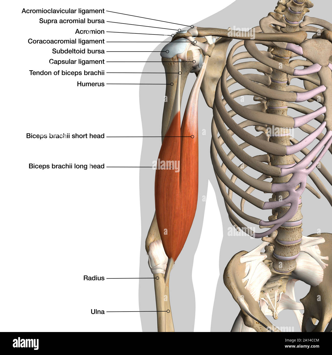
The muscles of the chest and upper back occupy the thoracic region of the body inferior to the neck and superior to the abdominal region and include the muscles of the shoulders. When you think of abs, what muscle do you typically think of? Plus, how to target each to make them bigger and stronger. This might sound like a strange question, right? Chest muscles function in respiration while abdominal muscles function in torso movement and in maintenance of balance and posture. Labeled muscles of lower leg. This mri chest (thorax) axial cross sectional anatomy tool is absolutely free to use. Here is the same image with the chest muscles labeled. Pectoralis major pectoralis minor serratus anterior subclavius Related to human arm muscles anatomy. The pectoralis major and the pectoralis minor, known collectively as your pecs. The pectoral region is located on the anterior chest wall. It contains four muscles that exert a force on the upper.
Here is the same image with the chest muscles labeled. But in actuality there are 4 separate muscles that contribute to your overall abdominal development. These important muscles control many motions that involve moving the arms and head — such as throwing a ball, looking up at the sky, and raising your hand. Here, we break down the anatomy of your chest muscles. Computed tomography (ct) of the chest can detect pathology that may not show up on a conventional chest radiograph(1).
Abdominal Muscles Diagram High Resolution Stock Photography And Images Alamy from c8.alamy.com Ct neck with annotated scrollable images. Supraspinatus, infraspinatus, subscapularis, and teres minor. The pectoral region is located on the anterior chest wall. The muscles of the chest and upper back occupy the thoracic region of the body inferior to the neck and superior to the abdominal region and include the muscles of the shoulders. Each of these muscles has its origin on the scapula and inserts around the head of the humerus. Related to human arm muscles anatomy. Coronal c+ portal venous phase. The pectoralis major and the pectoralis minor, known collectively as your pecs.
The chest or thorax is the region between the neck and diaphragm that encloses organs, such as the heart, lungs, esophagus, trachea, and thoracic diaphragm.
The anterior serratus pulls the scapula outward which lifts the shoulder. Ventral trunk muscles (overview) the trunk (torso) is the central part of the body to which the head and the limbs are attached. Its functions are to move the scapula forward and upward. Labeled muscles of lower leg. Muscles of the chest and their functions you have two mighty muscles on both sides of your chest: Muscles and layers of the thoracic cavity. The pectoralis major, pectoralis minor, serratus anterior and subclavius. Anatomy and physiology questions and answers. Pectoralis major pectoralis minor serratus anterior subclavius The torso muscles attach to the skeletal core of the trunk, and depending on their location are divided into two large groups: Plus there are links to lots of other great anatomy and physiology quizzes and other resources; The tendons of these muscles surround and support the humerus while the contraction of the muscles rotates, adducts, or abducts the humerus. Supraspinatus, infraspinatus, subscapularis, and teres minor.
The torso muscles attach to the skeletal core of the trunk, and depending on their location are divided into two large groups: Alles rund um kostüme & verkleiden. The muscles of the chest and upper back occupy the thoracic region of the body inferior to the neck and superior to the abdominal region and include the muscles of the shoulders. Except for the brain, the trunk houses all the vital organs of the human body. Male shoulder and chest muscles labeled chart on white labeled human anatomy diagram of male shoulder, biceps, arm, and chest muscles frontal anterior view on a white background.
Male Arm And Chest Muscles Labeled Chart On White Stock Photo Download Image Now Istock from media.istockphoto.com It also protects several vital organs of the chest, such as the heart, aorta, vena cava, and. Label muscles of the chest in figete 12.9.) e and deep muscles on the left side. Label muscles of the chest in figete 12.9.) e and deep muscles on the left side. Coronal c+ portal venous phase. Muscles of the chest and their functions you have two mighty muscles on both sides of your chest: Chest muscle anatomy the pectoralis major muscles also known as the pecs are located on the front of the rib cage and form the major muscles of the chest. Leg muscles anatomy leg anatomy human body anatomy human anatomy and physiology muscle anatomy anatomy study upper leg muscles thigh muscles chest muscles. I mean, the abs are the muscle.
This muscle group is responsible for pushing movements and interacts synergistically with the anterior deltoid of the shoulder and triceps of the arm.
The pectoralis major and the pectoralis minor, known collectively as your pecs. Ventral trunk muscles (overview) the trunk (torso) is the central part of the body to which the head and the limbs are attached. You go to the gym to train your abs. Muscles and layers of the thoracic cavity. But in actuality there are 4 separate muscles that contribute to your overall abdominal development. Muscles of the chest, also called the thorax, include both smooth muscles and skeletal muscles. Serratus anterior superior, serratus anterior intermediate, serratus anterior inferior and runs from the front of the chest around the side to the scapula. Plus there are links to lots of other great anatomy and physiology quizzes and other resources; Plus, how to target each to make them bigger and stronger. Its functions are to move the scapula forward and upward. Label muscles of the chest in figete 12.9.) e and deep muscles on the left side. This might sound like a strange question, right? This mri chest (thorax) axial cross sectional anatomy tool is absolutely free to use.
This mri chest (thorax) axial cross sectional anatomy tool is absolutely free to use chest muscles anatomy. Here is the same image with the chest muscles labeled.